DSPE-PEG-PDP辅助制备的柔性PEG垫层用于脂质双层膜重构研究
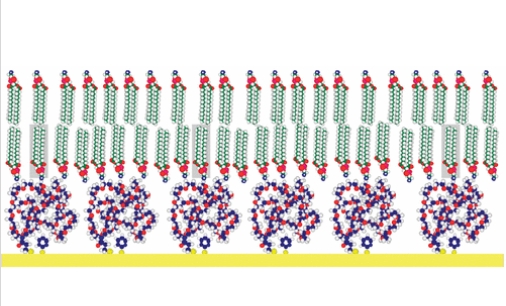
文献综述:聚乙二醇负载电阻脂质单层在金面的原位组成及定性分析超链接://pubs.acs.org/doi/abs/10.1021/la048378o写作者:杰弗里·C·芒罗,柯蒂斯·W·弗兰克文献综述:有玩家给出,在脂质两层膜和膏状表层直接增加整合物垫层,都可以建立挺括、可变气门正时形的膜层,而做到跨膜蛋白酶的插入表格和转化。本设计顺利在两步来完成过滤具体步骤,在聚乙二醇 (PEG) 形式上建立了可转化的、可束博的脂质两层膜。 PEG 胶片是顺利在共过滤异功能表表远爪 PEG 脂质整合物(1,2-二硬脂酰-sn-甘油-3-磷酸甲醇胺-N-聚(乙二醇)-2000- N- [3-(2-(吡啶基二硫代)丙酸酯])(DSPE-PEG-PDP)和非脂质功能表表化的 PEG-PDP 从甲醇/水混合型物中制作的,如已经的医学论文(Munro,JC;Frank,CW Langmuir 2004,20,3339-3349)相应述。第二选用三步脂质粘附物攻略 。第一步,将脂质从己烷溶剂中粘附物到 PEG 质粒上。此外,将囊泡粘附物并相融在表明上以在水的环境中变成多层高层。光固色工作后的荧光恢复正常揭示,该的过程 所产生扩撒标准值约为 2 μm /s。随束博脂质密度计算公式的新增,多层高层膜的迁徙率也随之大幅度降低。表明上等化合物体共震法(适用法测定法原位膜重量)和荧光法(适用化学发光法法测定法每项18 x 18豪米样件的荧光标准)也断定了多层高层膜的变成,在于多层高层型式。可惜的是,荧光显微镜排查也界面显示样件上出现相对较大的缺点,这局限了该体统的使用性。Abstract ImageInclusion of a polymer cushion between a lipid bilayer membrane and a solid surface has been suggested as a means to provide a soft, deformable layer that will allow for transmembrane protein insertion and mobility. In this study, mobile, tethered lipid bilayers were formed on a poly(ethylene glycol) (PEG) support via a two-step adsorption process. The PEG films were prepared by coadsorbing a heterofunctional, telechelic PEG lipopolymer (1,2-distearoyl-sn-glycero-3-phosphoethanolamine-N-poly(ethylene glycol)-2000-N-[3-(2-(pyridyldithio)propionate]) (DSPE-PEG-PDP) and a nonlipid functionalized PEG-PDP from an ethanol/water mixture, as described in a previous paper (Munro, J. C.; Frank, C. W. Langmuir2004, 20, 3339−3349). Then a two-step lipid adsorption strategy was used. First, lipids were adsorbed onto the PEG support from a hexane solution. Second, vesicles were adsorbed and fused on the surface to create a bilayer in an aqueous environment. Fluorescence recovery after photobleaching experiments show that this process results in mobile bilayers with diffusion coefficients on the order of 2 μm2/s. The mobility of the bilayers is decreased slightly by increasing the density of tethered lipids. The formation of bilayers, and not multilayer structures, is also confirmed by surface plasmon resonance, which was used to determine in situ film thickness, and by fluorimetry, which was used to determine quantitatively the fluorescence intensity for each 18 by 18 mm sample. Unfortunately, fluorescence microscopy also shows that there are large defects on the samples, which limits the utility of this system.


 杏彩体育平台
微信公众号
 官方微信
官方微信